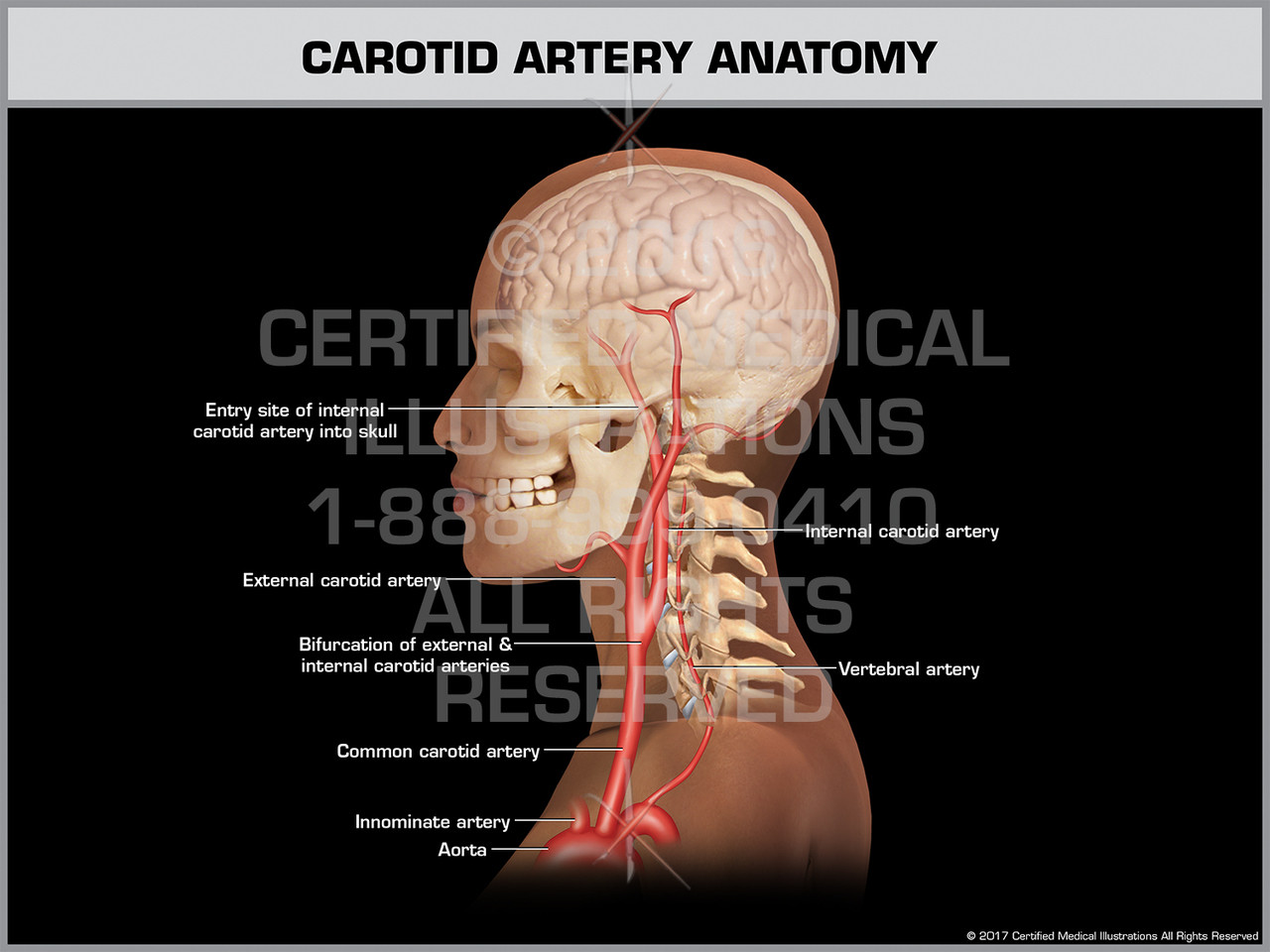
Anatomy Arteries In Neck / Examination Of The Neck Veins Nejm / In human anatomy, they arise from the common carotid arteries where these bifurcate into the internal and external carotid arteries at cervical vertebral level 3 or 4.
Anatomy Arteries In Neck / Examination Of The Neck Veins Nejm / In human anatomy, they arise from the common carotid arteries where these bifurcate into the internal and external carotid arteries at cervical vertebral level 3 or 4.. Occipital artery internal carotid artery external carotid artery vertebral artery subclavian artery. This section of the website will explain large and minute details of arterial anatomy of neck. The arteries are attached to the deep layers of the dermis, and profuse bleeding that is commonly seen in scalp wounds is attributed to a relative inability of the arteries to retract due blood supply of the scalpthe arterial blood supply to the scalp is via the internal and external carotid artery system. Illustration of the arterial system in the human body, shown in a standing figure. Neck, in land vertebrates, the portion of the body joining the head to the shoulders and chest.
Anatomy of the neck head anatomy body anatomy arteries anatomy. The internal carotid artery (latin: Carotid artery ijv sympathetic chain cn ix, x, xi, xii. Arteria carotis interna) is located in the inner side of the neck in contrast to the external carotid artery. The neck is the area between the skull base and the clavicles.
Anatomy Of Carotid Artery Anatomy Drawing Diagram from ars.els-cdn.com The right and left subclavian arteries give rise to the thyrocervical trunk. Cca typically divides at the level of c3 or c4 vertebral. Part of the occipital and parts of the transverse cervical and suprascapular arteries are also found in the occipital triangle. Learn about the types of arteries and how they function. Thus the vascular territory supplied by the striate arteries is particularly susceptible to lacunar infarcts. The head rests on the top part of the vertebral column, with the skull joining at c1. The head and neck receives the majority of its blood supply through the carotid and vertebral arteries. Arteries of the head and neck.
.veins and arteries of the neck activate javascript arteries in the neck diagram, common carotid artery branches, external carotid artery function, how many carotid arteries, left common carotid artery function, the left common carotid artery supplies blood to the, what does the external carotid artery …
The head rests on the top part of the vertebral column, with the skull joining at c1. Arteria carotis interna) is located in the inner side of the neck in contrast to the external carotid artery. .veins and arteries of the neck activate javascript arteries in the neck diagram, common carotid artery branches, external carotid artery function, how many carotid arteries, left common carotid artery function, the left common carotid artery supplies blood to the, what does the external carotid artery … Overview of arteries of the neck 0:16. Vagus nerve or sympathetic plexus: Cca typically divides at the level of c3 or c4 vertebral. An artery is an elastic blood vessel that transports blood away from the heart. The neck is the area between the skull base and the clavicles. Create flashcards for free and quiz yourself with an interactive flipper. Head and neck anatomy quiz 2: Instant anatomy is a specialised web site for you to learn all about human anatomy of the body with diagrams, podcasts and revision questions. Through its external carotid branch, it supplies the face, scalp, tongue, upper and lower teeth, gums, sinus, external and middle ear, pharynx and larynx in the throat, as well as the thyroid. Want to learn more about it?
The common carotid artery is a primary source of oxygenated blood to the head and neck. Hollow spaces within the cervical vertebrae protect and conduct the spinal cord and vertebral arteries through the neck. The 5 anatomical spaces of the infrahyoid neck. The anatomy of neck arteries, normal variations, and anastomoses between different arteries is discussed in this chapter. The neck is the area between the skull base and the clavicles.
Carotid Artery Anatomy from cdn10.bigcommerce.com Create flashcards for free and quiz yourself with an interactive flipper. Anatomy of the neck head anatomy body anatomy arteries anatomy. 17 933 просмотра 17 тыс. An artery is an elastic blood vessel that transports blood away from the heart. The external carotid artery stretches upwards from the level of upper border of the lamina of the thyroid cartilage to a stage supporting the neck of the mandible, where it ends in the substance of the parotid gland by splitting. 19 heart & neck vessels flashcards. In radiology, the 'head and neck' refers to all the anatomical structures in this region excluding the central nervous system, that is, the brain and spinal co. Narrowing of the arteries, usually caused by atherosclerosis.
Vertebral arteries (arteria vertebralis) next to your spine which become basilar artery (arteria basillaris) and brings blood to the back of the brain.
The internal carotid artery (latin: Cca typically divides at the level of c3 or c4 vertebral. Immediate treatment is necessary to restore blood flow in the artery. The head rests on the top part of the vertebral column, with the skull joining at c1. The carotid sheath plays an important role in head and neck anatomy and contains several vital structures, including the carotid artery, jugular vein a carotid endarterectomy is performed to excise atherosclerotic thickening of the intima within the internal carotid artery in an effort to reduce strokes. The head and neck receives the majority of its blood supply through the carotid and vertebral arteries. The 5 anatomical spaces of the infrahyoid neck. A sudden blood clot in one of the arteries, stopping blood flow. Neck, in land vertebrates, the portion of the body joining the head to the shoulders and chest. The common carotid artery is a primary source of oxygenated blood to the head and neck. Vagus nerve or sympathetic plexus: The cca courses superiorly in the neck, anteromedial to the jugular vein and alongside the vagus nerve. Head and neck anatomy quiz 2:
Note the feathery network of blood vessels in the left and right lungs (next to the heart). The external carotid artery stretches upwards from the level of upper border of the lamina of the thyroid cartilage to a stage supporting the neck of the mandible, where it ends in the substance of the parotid gland by splitting. Narrowing of the arteries, usually caused by atherosclerosis. Hollow spaces within the cervical vertebrae protect and conduct the spinal cord and vertebral arteries through the neck. Despite being a relatively small region, it contains a range of important anatomical features.
Vertebral Artery Course Segments Branches Kenhub from thumbor.kenhub.com Despite being a relatively small region, it contains a range of important anatomical features. The mass is not in between the. Medial displacement of lateral documents similar to basic anatomy of the neck. The arteries are attached to the deep layers of the dermis, and profuse bleeding that is commonly seen in scalp wounds is attributed to a relative inability of the arteries to retract due blood supply of the scalpthe arterial blood supply to the scalp is via the internal and external carotid artery system. The portal vein is typically formed by the union of the superior mesenteric and the splenic veins posterior to the neck of the pancreas. Arteries of the head and neck. A blocked artery in neck may lead to excessive pain in jaw / face may lead to death of that unlucky person if not attended to by medical experts. The right and left subclavian arteries give rise to the thyrocervical trunk.
Carotid artery ijv sympathetic chain cn ix, x, xi, xii.
The arteries are attached to the deep layers of the dermis, and profuse bleeding that is commonly seen in scalp wounds is attributed to a relative inability of the arteries to retract due blood supply of the scalpthe arterial blood supply to the scalp is via the internal and external carotid artery system. The neck is the area between the skull base and the clavicles. 17 933 просмотра 17 тыс. Learn about the types of arteries and how they function. 19 heart & neck vessels flashcards. Vertebral arteries (arteria vertebralis) next to your spine which become basilar artery (arteria basillaris) and brings blood to the back of the brain. Carotid artery ijv sympathetic chain cn ix, x, xi, xii. Hollow spaces within the cervical vertebrae protect and conduct the spinal cord and vertebral arteries through the neck. Some important structures contained in or passing through the neck include the seven cervical vertebrae and enclosed spinal cord, the jugular veins and carotid arteries, part of the esophagus, the larynx. Immediate treatment is necessary to restore blood flow in the artery. 15.16 renal veins anterior view. Instant anatomy is a specialised web site for you to learn all about human anatomy of the body with diagrams, podcasts and revision questions. Which artery does not supply blood to t… which of the following are branches of…
The common carotid artery is a primary source of oxygenated blood to the head and neck arteries in neck. The common carotid artery is a primary source of oxygenated blood to the head and neck.


0 Komentar